Pulsed waves transmitted at constant pulse repetition frequency and only one sample as function of time is collected 3. The present invention provides a kind of power doppler blood flow composite imaging method and system and its method comprises the steps.

Color Doppler Fujifilm Visualsonics
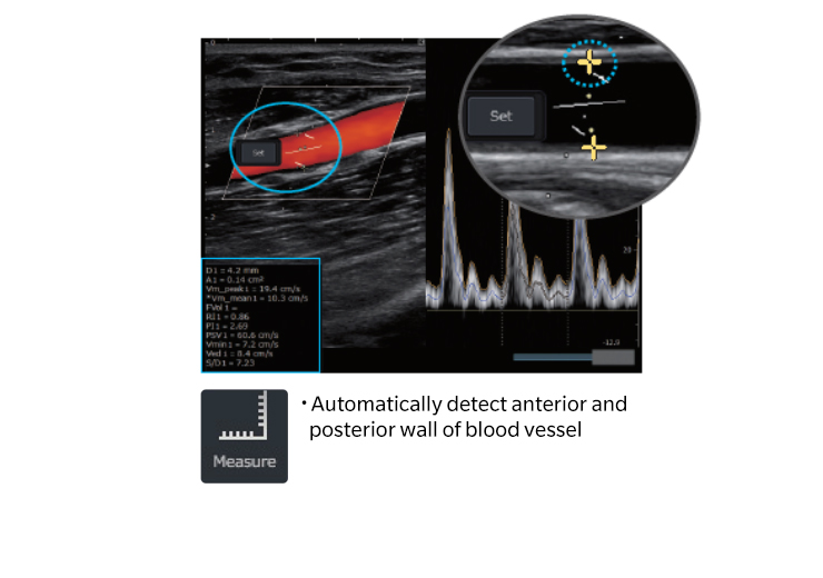
In accordance with either a blood flow velocity or blood flow energy or both of them of each pixel point in a blood flow image determining suspected blood flow boundary points.

. With respect to the method of blood flow measurement there are two kinds. Continuous sinusoidal wave transmitted with one crystal and reflected wave received with second crystal 2. A nuclear heart scan is an imaging test that uses special cameras and a radioactive substance called a tracer to create pictures of your heart.
Ultrasound is composed of sound waves with frequencies which are significantly higher than the range of human hearing 20000 Hz. The test can detect problems with blood flow in the heart tissue and can diagnose ischemic heart disease. A Doppler ultrasound study a technique that evaluates blood flow through a blood vessel is usually part of this exam.
Color Flow CF imaging. A penile Doppler test is used to determine blood flow within a persons erect. Ultrasound is not different from normal audible sound in its physical properties except that.
With the sonographic technique of color Doppler flow imaging CDFI the spatial and temporal distribution of the color-coded Doppler signal can be visualized in realtime and superimposed on a high-resolution gray-scale image of tissue and vessel morphology. Pulsed Wave PW Doppler. In June 2016 undertook the Special Diagnosis and Treatment RD Major Project of National Ministry of Science and Technology -Fusion imaging of noninvasive vascular elasticity and vector blood flow and its implementation in portable ultrasound diagnostic units of China.
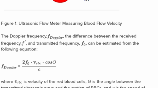
Ultrasound image sonogram of a fetus in the womb viewed at 12 weeks of pregnancy bidimensional scan An ultrasonic examination. In the meantime the feasibility of measuring blood flow in the heart and vessels using the Doppler effect in ultra- sonic waves has become well known. This flow meter uses Doppler technology and the main intention of this meter is to deliver the analysis of blood flow.
Ultrasound is sound waves with frequencies higher than the upper audible limit of human hearing. The method comprises the following steps. The first ultrasonic flow meter was invented by Japanese physicist namely Shiego Satomura in the year 1959.
Depending on the number of the suspected blood flow boundary points of each. Functional ultrasound applications include Doppler and color Doppler ultrasound for measuring and visualizing blood flow in vessels within the body or in the heart. Some were as large as a room.
At present there are many manufacturing companies were designing different types of clamp-on flow. The middle hepatic vein provides a land-mark to. During that time ultrasound waves are directed toward the area of interest.
Over the past twenty-five years many methodological approaches have been developed to provide an assessment of endothelial function in humans. DOPPLER IMAGING It is a general term used to visualize velocities of moving tissues. Ultrasonic images also known as sonograms are created by sending pulses of ultrasound into tissue using a probe.
These relax the corpora cavernosa to the extent that help develop erections faster and. Ultrasonic blood flowmeter An ultrasonic flow meter is a type of flow meter that measures the velocity of a fluid with ultrasound to calculate volume flow. When microbubbles in the blood flow pass the imaging window the microbubbles compressible gas cores oscillate in response to the high frequency sonic energy field.
Blood flow may be conventionally visualized via high frequency ultrasound power Doppler imaging. Simple 2D imaging devices developed by researcher in US and Japan. In addition to imaging the arterial walls the ultrasound techniques can measure the blood flow velocity by making use of the Doppler effectThe reflected ultrasound is shifted in frequency from the frequency of the source and that difference in frequency.
Due to transmitted signals of multi-cycled burst with an array imaging system this may require high frequency transmission over 70 MHz to reach the required fine spatiotemporal resolution. Recent advances in imaging technology offer the user the ability to evaluate the portal venous system FIG. The profession sonographer was created by the AMA.
It can also assess how. It can also measure the speed of the blood flow and direction of movement. Doppler ultrasound evaluates blood velocity as it flows through a blood vessel.
Waveform image bottom right shows the sound of flowing blood in the carotid artery. Continuous wave Doppler and pulse wave Doppler or the so-called pulsed Doppler. Incidentally this is the same principle that erection pills like Viagra use.
Blood flow through the heart and large vessels has certain characteristics that can be measured using Doppler instruments. After four years the earliest flow meters have appeared in industrial applications. Doppler measurements of tissue motion and blood flow Japan US.
To configure some scanning lines in same frame scanning and is that every scanning line configures certain scanning direction wherein and the number N 2 in described scanning directionScanning line wave beam is launched to blood flow to be imaged. Continuous Wave CW Doppler. The microbubbles will remain in the systemic circulation for a certain period of time.
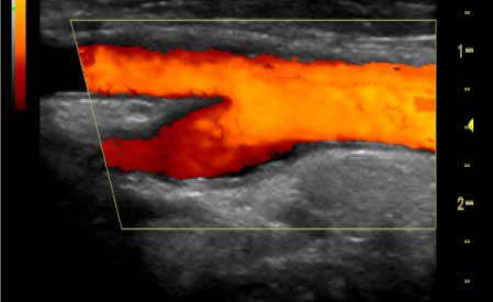
A Doppler ultrasound is a non-invasive method that uses high-frequency sound waves or ultrasound reflected off circulating red blood cells to estimate the blood flow through your blood vessels. Transverse color Doppler image shows flow in the left middle and right hepatic veins draining into the inferior vena cava IVC. The speed of blood flow is assessed by measuring the rate of change in the frequency of the reflected sound waves.
The invention provides an ultrasonic color blood flow imaging boundary processing method and system. Ultrasound of carotid artery. Using the general principle of imaging that you cant see anything smaller than the wavelength suggests a 02 mm ultimate resolution limit.
A few studies using CDFI have examined flow patterns in the. Using ultrasonic transducers the flow meter can measure the average velocity along the path of an emitted beam of ultrasound. If you could increase the amount of blood that flows into the cavernosa tissues then you could get better erections and perhaps for longer periods of time than before.
Introduced in 1989 the FMD test incorporates a forearm occlusion and subsequent reactive hyperemia that promotes nitric oxide production and vasodilation of the brachial artery. Ultrasound does not use ionizing radiation has no known harmful effects and provides images of soft tissues that dont show up on x-ray images. Its ultimately the blood flow to your penis that decides the quality of your erections.
Vascular Navi Ultrasound Healthcare Konica Minolta
Using Ultrasounds To Measure Blood Flow Velocity Practice Khan Academy
Healthy Aging And The Blood Brain Barrier Nature Aging

Pin On Pinboard Highlighting Our Recent Publications

Fluid Aspirate Color Characteristics Note Anchovy Sauce Appearance Amoebic Liver Abscess Medical Studies Surgery Diagnosis

Propagation Speed Error Artifacts Ultrasound Physics Ultrasound Sonography Ultrasound

Behavioral Neuroscience Fear Circuitry Behavioral Neuroscience Chronic Fatigue Symptoms Fibromyalgia Causes

Micturating Cystourethrography Mcu Vcug Bladder Catheter Medical Assistant
![]()
Ultrasonic Piezo Transducer For Blood Flow Monitoring

Bulbar Pseudobulbar Palsy Goh Hk
Color Doppler Fujifilm Visualsonics

Pin On Embedded Articles Projects

Pin On Clinical
![]()
Ultrasonic Piezo Transducer For Blood Flow Monitoring

Windword Oil On Canvas Visionary Art Art
Vascular Ultrasound

Bulbar Pseudobulbar Palsy Goh Hk

Zbd99272l9nzxm

Pin By Nonas Arc On Right Ventricular Outflow Tract Obstruction Atrial Septal Defect Stenosis Tetralogy


0 komentar:
Posting Komentar